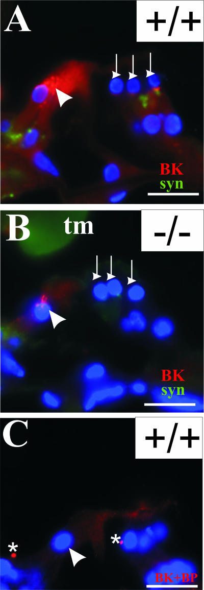
Figure 8.
Reduced BK α-subunit expression in the organ of Corti of Tmprss1−/− mice. BK α-subunit (red) and synaptophysin (green) expression in the organ of Corti of adult wild-type (A) and age-matched Tmprss1−/− (B) mice. C: The BK α-subunit antibody was preincubated with the corresponding anti-peptide before being applied to sections. White arrowhead indicates the upper part of inner hair cells. White downward arrows point to the base of outer hair cells. Asterisks indicate background fluorescence, which does not localize within any cellular compartments. Cell nuclei are labeled with 4′,6-diamidino-2-phenylindole dihydrochloride (blue). Scale bar = 20 μm.

