Figure 5.
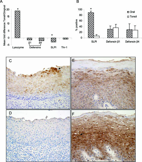
SLPI expression in the tonsil and oral epithelia. A: Mean fold change of gene expression between tonsil and gingiva for the innate immune factors lysozyme, defensin-β1, defensin-β4, SLPI, and thrombospondin-1. *P ≤ 0.05. B: Quantitation of percent positive SLPI, defensin-β1, and defensin-β4 cells in the tonsil (n = 5) and oral epithelium (n = 5). *P ≤ 0.05. C: SLPI staining in the tonsil is weak (grade 1) with isolated cells staining moderately (grade 2, arrow showing higher intensity staining). D: Negative control. In the oral mucosa, SLPI staining is abundant and of high intensity (grade 2 to 3) for both keratinized (E) and nonkeratinized (F) oral epithelia (arrow shows high-intensity staining). Original magnifications: ×20 (D–F).

