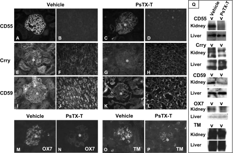
Figure 3.
Expression of CReg 24 hours after PsTX-T administration under Immunofluorescence and Western blot analyses. Sections were stained for CD55 (A–D), Crry (E–H), CD59 (I–L), Thy 1.1 antigen (M and N), or thrombomodulin (O and P). A, B, E, F, I, J, M, and N show sections from rats injected with vehicle, whereas C, D, G, H, K, L, O, and P are from rats injected with PsTX-T. Exposure times and other parameters were kept constant to permit comparison. The expression of CD55, Crry, and CD59 was clearly decreased in glomeruli after PsTX-T exposure. In contrast, the expression of Crry and CD59 in tubular epithelium was preserved except where the epithelium was damaged (H and L). Original magnifications: ×400 (A, C, E, G, I, K, and M–P); ×200 (B, D, F, H, J, N, and O). Asterisk, glomerulus. TM, thrombomodulin. Q: Results of Western blot analysis. The left of each blotting set was the lysate from rats injected with PsTX-T, and the right was from rats injected with vehicle. The samples and the probed antigens are displayed at the left.

