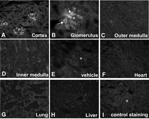
Figure 5.
The tissue distribution of PsTX-T after intravenous administration. Intravenously administered PsTX-T was detected using specific antiserum in glomerular endothelium (A and B) 10 minutes after injection. At this time, there was no specific staining in the interstitium of cortex (A), in outer medulla (C), or in inner medulla (D). There was also no detectable staining in heart (F), lung (G), and liver (H) after administration of toxin. Asterisks in A, E, and I show positions of glomeruli. E: Anti-PsTX-T staining in kidney with vehicle administration instead of PsTX-T. I: Staining of PsTX-T-treated kidney with nonimmune mouse serum. Original magnifications: ×200 (A and C–H); ×400 (B).

