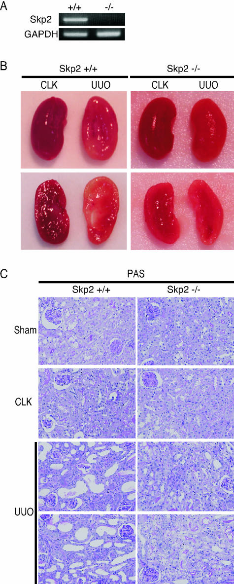
Figure 3.
Levels of Skp2 and GAPDH mRNA expression (A) and the representative macroscopic (B) and microscopic (C; periodic acid-Schiff, magnification, ×300) findings of the obstructed (UUO) and nonobstructed contralateral (CLK) kidneys of Skp2+/+ (left) and Skp2−/− mice (right) 7 days after UUO. No detectable expression of Skp2 mRNA was observed in Skp2−/− mice (A). Although remarkable renal atrophy was noted in the obstructed kidneys from Skp2+/+ mice, it was markedly less in Skp2−/− mice (B). Tubular dilatation and atrophy and interstitial cell infiltration were observed in obstructed kidneys from Skp2+/+ mice. However, the severity of these lesions was markedly less in obstructed kidneys from Skp2−/− mice (C).

