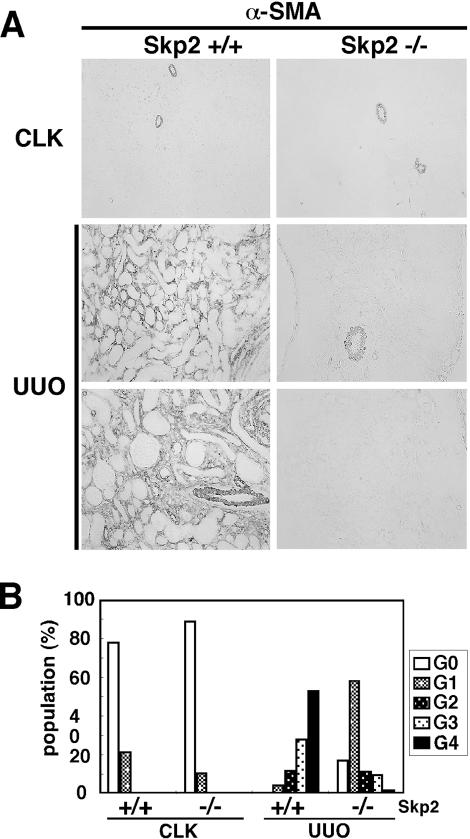
Figure 8.
Representative pictures of the CLK and UUO kidney sections from Skp2+/+ mice (A, left) and Skp2−/− mice (A, right) and the semiquantification of immunostaining intensity (B) for α-SMA 7 days after obstruction. Significant increases of interstitial myofibroblastic cells positive for α-SMA were observed in obstructed kidneys from Skp2+/+ mice on day 7, but the levels were markedly attenuated in obstructed kidneys from Skp2−/− mice (A, magnification, ×300). The grading of immunostaining intensities for α-SMA shown in B was as follows: G0 (grade 0, positive area 0%); G1 (grade 1, ≤20% in interstitial area were positive); G2 (grade 2, 21 ≈ 50% positive); G3 (grade 3, 51 ≈ 80% positive); and G4 (grade 4, ≥81% positive). P < 0.0001; Skp2+/+ UUO versus Skp2−/− UUO.

