
Figure 5. Analysis of potential involvement of PAF receptor in OxPAPC-induced signaling and endothelial barrier protection.
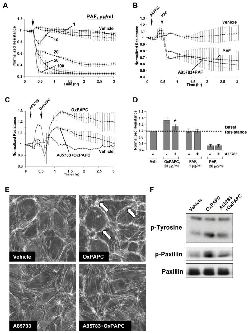
Panels A – D: TER measurements in EC monolayers. Cells grown on gold microelectrodes were stimulated with PAF (1 μg/ml, 5 μg/ml, 20 μg/ml, 50 μg/ml, 100 μg/ml) (panel A); or were preincubated with PAF receptor antagonist A85783 (5 μM, 30 min) followed by stimulation with 20 μg/ml of PAF (panel B) or OxPAPC (panel C). Effects of PAF receptor antagonist on PAF- and OxPAPC-mediated permeability changes are summarized in panel D. Shown are pooled data of three independent experiments represented as mean + SD. *p<0.01 vs OxPAPC alone. Panel E: EC were preincubated with A85783 (5 μM, 30 min) followed by stimulation with OxPAPC (20 μg/ml, 30 min) and immunofluorescent analysis of F-actin remodeling by staining with Texas Red phalloidin. Panel F: EC preincubated with vehicle or A85783 (5 μM, 30 min) were treated with OxPAPC, and total protein tyrosine phosphorylation and site-specific paxillin phosphorylation was detected by immunoblotting with phospho-tyrosine or phospho-paxillin (Tyr118) antibodies. Equal protein loading was confirmed by reprobing the membranes with paxillin antibody.