
Figure 1. The phenotype and genotype of CML.
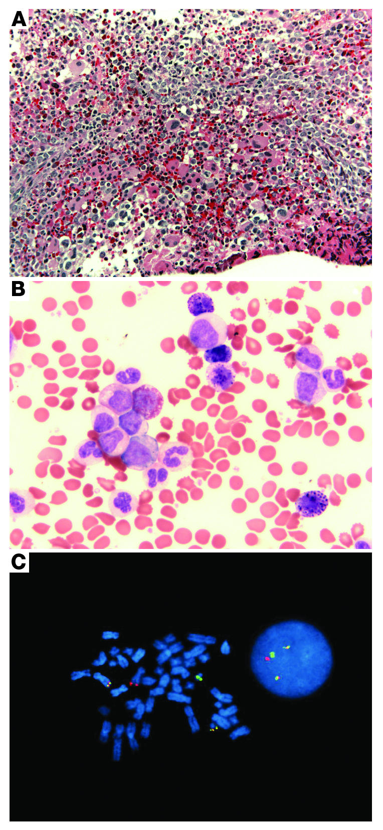
(A) A bone marrow biopsy from a patient with CML shows the typical hypercellularity with granulocytic and megakaryocytic hyperplasia (original magnification, ×200). (B) The peripheral blood is characterized by a full spectrum of myeloid cells, including immature myeloid cells with rare blasts. Basophilia is also observed (original magnification, ×630). (C) Dual-color, dual-fusion FISH displaying BCR-ABL signals in bone marrow cells in metaphase (left) and interphase (right). The red fluorescent probe is specific for ABL, while the green probe is specific for BCR. Yellow signals the presence of BCR-ABL and ABL-BCR fusions.