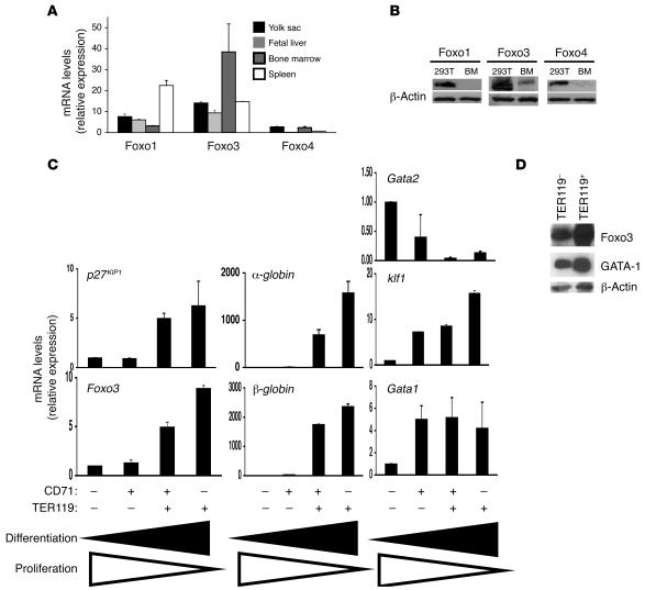
Figure 4. Foxo3 expression is upregulated during erythroblast maturation.
(A) QRT-PCR analysis of Foxo1, Foxo3, and Foxo4 in embryonic and adult hematopoietic organs. Shown are representative results from 3 independent experiments performed in duplicate. (B) Western blot analysis of endogenous FoxO protein expression in the bone marrow. Protein lysates of HEK293T cells overexpressing FOXO1, FOXO3a, and FOXO4 cDNAs were used as positive controls. (C) QRT-PCR analysis of indicated transcripts from subpopulations of fetal liver isolated by flow cytometry according to their CD71 and TER119 expression. Note that expression of Foxo3 is the highest in the most mature erythroid cell subpopulation (CD71–TER119+). Representative results from 3 independent experiments performed in triplicate are shown as mean ± SEM. Cells differentiate from a double CD71–TER119– cell subpopulation enriched in hematopoietic progenitors, including erythroid progenitors, to CD71+TER119– cells containing mostly proerythroblasts and basophilic erythroblasts, then to CD71+TER119+ cells enriched for basophilic/polychromatophilic erythroblasts, and finally to CD71–TER119+ cells consisting mostly of normoblasts. (D) Western blot analysis of Foxo3 in subpopulations of fetal liver enriched for progenitors (TER119–) and for erythroid precursors (TER119+, erythroblasts) using anti-FOXO3a antibody. Anti–GATA-1 (N6) and anti–β-actin antibodies were used as controls.

