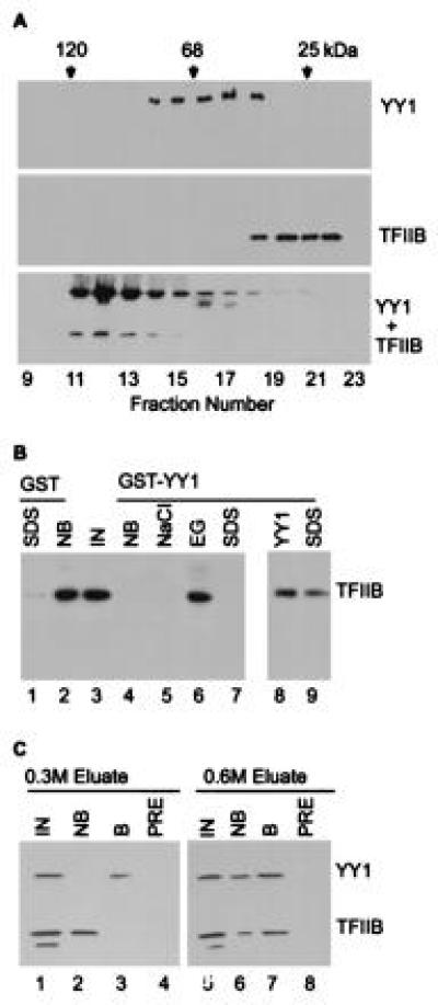
Figure 1.

TFIIB binds to YY1. (A) TFIIB binds to YY1 in solution. Purified TFIIB and YY1 were subjected to gel filtration chromatography alone or after mixing, and localized in the elution profile by protein blot assay using polyclonal antibody (18) to YY1 (Top), TFIIB (Middle), or a mixture of the antibodies (Bottom). Chymotrypsinogen A (25 kDa), BSA (68 kDa), and β-galactosidase (120 kDa) were markers. (B) TFIIB binds to GST–YY1. TFIIB was monitored by protein blot assay. Lanes: IN, TFIIB input to each reaction; NB, TFIIB not bound; NaCl, TFIIB eluted in buffer containing 1.0 M NaCl; EG, TFIIB eluted in buffer containing 50% ethylene glycol plus 100 mM NaCl; SDS, TFIIB eluted by boiling in buffer with detergent, either immediately after application to the affinity matrix (lanes 1 and 9) or following elution with ethylene glycol (lane 7); YY1, TFIIB eluted in buffer containing 60 μg/ml YY1. (C) Copurification of TFIIB and YY1 from a HeLa cell nuclear extract. Two fractions from a single-stranded DNA cellulose column (0.3 M and 0.6 M NaCl) containing both YY1 and TFIIB were assayed on a YY1-specific IgG matrix. TFIIB and YY1 were detected by protein blot assay. Lanes: IN, TFIIB and YY1 input to each reaction; NB, input which is not bound; B, input which is bound; PRE, input bound by preimmune IgG. Figures were produced using photoshop and freehand software.
