
Figure 2.
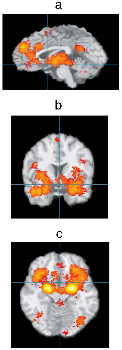
Preliminary summary of neuroimaging studies of core affective and emotion experiences. Activation foci were registered to a common stereotaxic brain atlas (Talairach & Tournoux 1988) where x = distance in millimeters to the right (+) or left (−) of midline; y = distance anterior (+) or posterior (−) to the anterior commissure; and z = distance superior (+) or inferior (−) to a horizontal plane through the anterior and posterior commissures. Midsagital (a, x = 0), coronal (b, y = 7), and horizontal (c, z = −13) images are presented. Significant areas of activation include OFC, insula, amygdala, ACC, and DMPFC (as well as VLPFC; not shown). VMPFC activations were also observed, but it is not clear that they extend down to the ventral surface, c, probably owing to problems with imaging that area of the brain. Lighter colors indicate a larger number of studies reported significant peak activations at that location (summary corrected for false discovery rate).