
Figure 2.
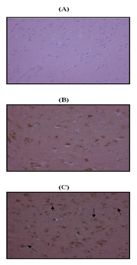
Immunohistochemical staining. ‘A’ represents the negative control using rabbit IgG. ‘B’ and ‘C’ are representative micrographs of immunohistochemistry obtained with a polyclonal antibody for 3-nitrotyrosine in control hippocampus and MCI hippocampus, respectively (×20 magnification). Intense nitrotyrosine staining is present in MCI hippocampus, whereas staining is far less prominent in control hippocampus. Nitrotyrosine is localized predominantly in neurons (arrows).