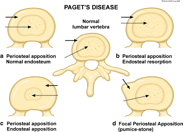
Fig. 1.

Diagram depicting the osseous mechanisms involved in vertebral body enlargement in Paget’s disease and its effect on the size of the marrow (dashed arrows) and cortex (solid arrows). A normal vertebra is depicted in the centre of the figure. a Periosteal apposition, normal endosteum resulting in thickened cortex, but with normal marrow size. b Periosteal apposition, endosteal resorption results in normal cortical thickness and an increased marrow size. c Periosteal apposition/endosteal apposition results in a thickened cortex and reduced marrow size. d Focal periosteal apposition results in a focal “pumice stone”-like enlargement
