
Fig. 20.
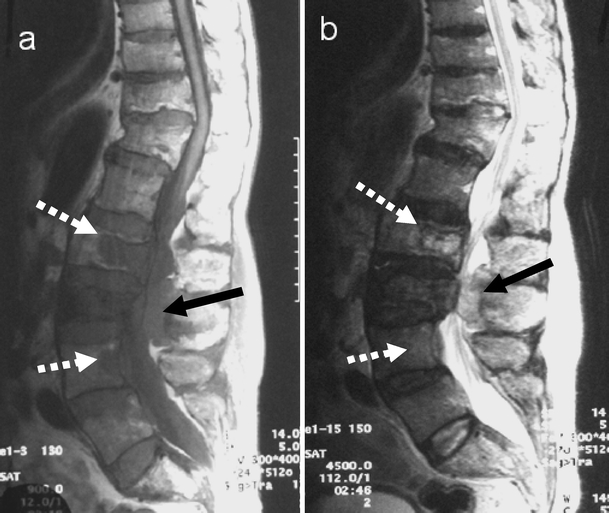
Vertebral metastasis from colon carcinoma. Sagittal a T1- and b T2-weighted MR images demonstrate metastasis in L2 and L4 vertebral bodies seen as discrete lesions (white arrows) with low signal on T1- and high signal on T2-weighted images. A further epidural lesion (black arrow) is seen in the spinal canal posteriorly at L3. Note the pagetic changes with expansion of L2 and L3 vertebral bodies