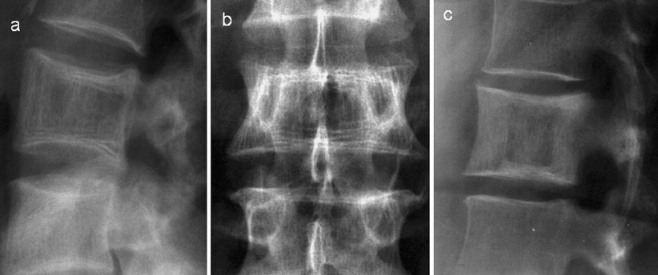
Fig. 6.

a Lateral and b antero-posterior radiographs demonstrate expansion of the vertebra with characteristic sclerotic lines parallel to the end-plates due to trabecular hypertrophy, an “early” sign of PD. c Lateral radiograph in a different patient demonstrates the “picture frame” vertebra due to thickening of the cortex and trabecular hypertrophy at the end-plates
