
Figure 4. Altered AQP expression in HCC is not unique to the H4IIE HCC model.
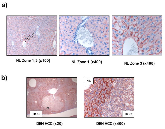
(a). Representative immunohistochemical images of normal mouse liver tissue probed with an antibody specific against AQP 9 (x100 magnification). Approximate representative hepatic lobule zones are labeled Z1, Z2 and Z3 (left panel). Increased magnification (x400) of zones 1 and 3 demonstrate increased Z3 (right panel) staining versus Z1 (center panel). (b) Representative immunohistochemical images of HCC tissue and surrounding normal liver tissue from a mouse-DEN model of HCC probed with anti-AQP 9 antibody (x20 and x400 magnification).