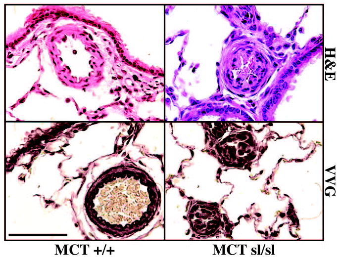
Figure 1.

Hematoxylin and eosin (H&E) staining of MCT+/+ rat lung revealed medial hypertrophy of small pulmonary arteries. VVG staining revealed a well-defined internal elastic lamina. In contrast, near occlusion of small pulmonary arteries in the MCTsl/sl rat lung is shown. VVG staining of MCTsl/sl rat lung of plexogenic lesions failed to reveal a well-defined internal elastic lamina. Bar=50 μm.
