
Figure 4.
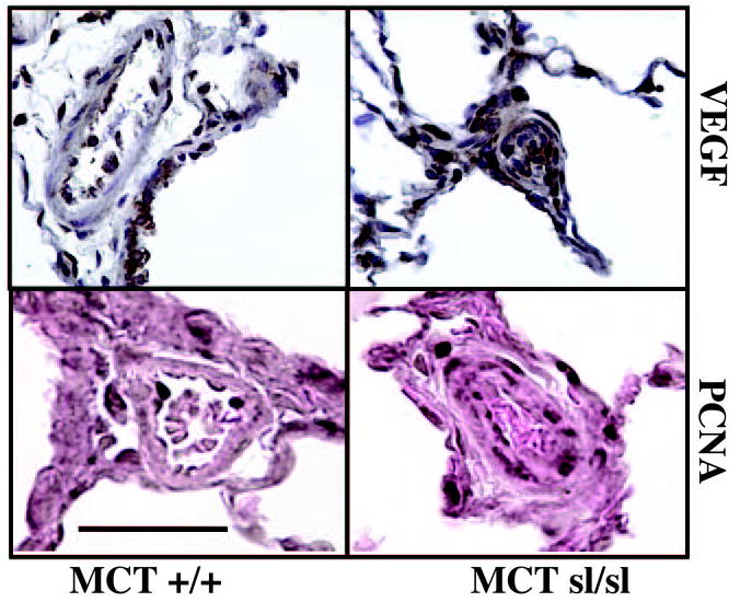
VEGF is expressed in alveoli and blood vessel wall of MCT+/+ rat lung. In plexogenic lesions in MCTsl/sl lungs, staining of the proangiogenic factor VEGF is noted. Immunohistochemical staining of MCT+/+ and MCTsl/sl rat lung for PCNA revealed PCNA-positive cells in EC layer of MCT+/+ group, whereas distribution of PCNA-positive cells in MCTsl/sl group was more widespread, being detected in the adventitial cell, medial cell, and EC layers. Bar=50 μm.