
Figure 2.
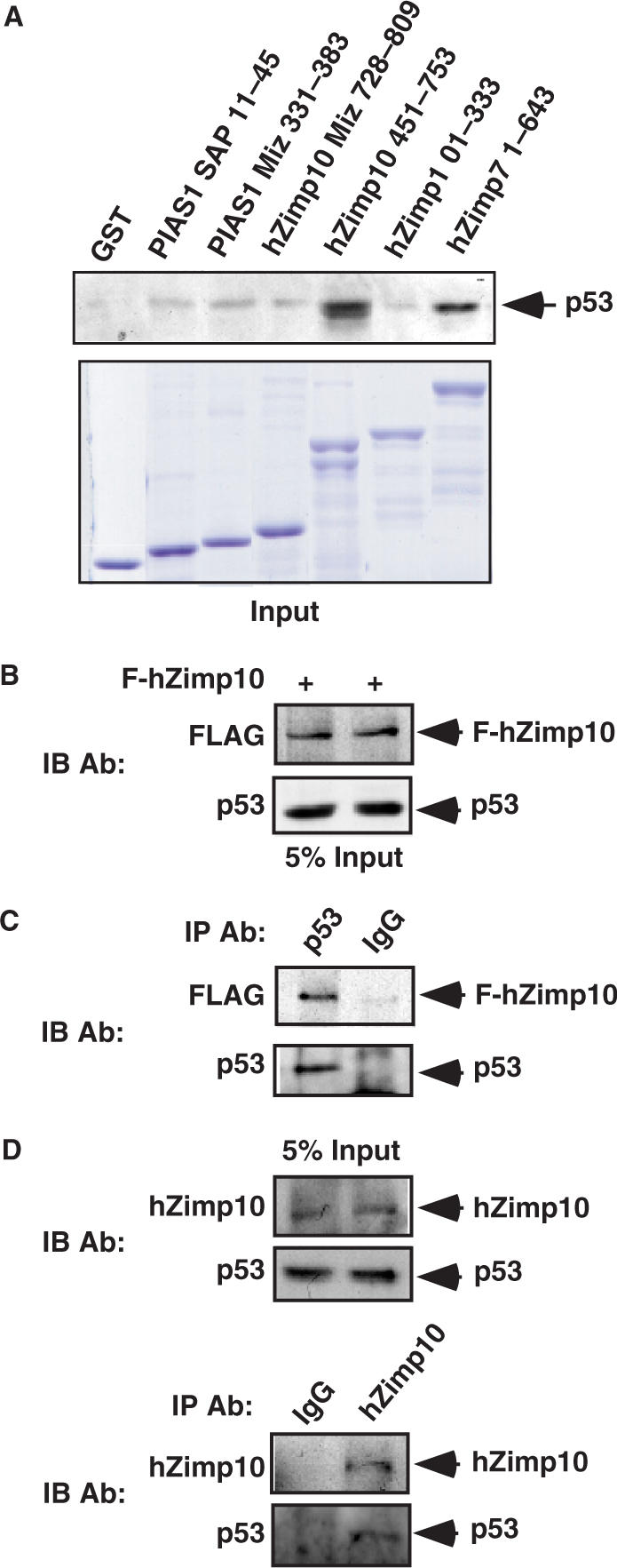
Physical interaction between hZimp10 and p53 in vitro and in intact cells. (A) Equal amounts of GST-PIAS1 MIZ domain, GST-PIAS1 SAP domain, GST-hZimp10 MIZ (728–809 amino acids), GST-hZimp10 Central (451–753 amino acids), GST-hZimp10 N-terminal (1–333 amino acids) and hZimp7 N-terminal (1–643 amino acids) fusion proteins were used to pull down endogenous p53 in MCF7 cells. GST protein alone was used as a negative control. Equal amounts of the above GST proteins were analyzed on SDS-PAGE. Material bound to GST columns was subjected to SDS-PAGE and western blot with p53-specific antibody. (B) HEK293 cells were transfected with pcDNA3-HA-p53 (0.1 μg) and pcDNA3-FLAG-tagged hZimp10 (1.8 μg). Here, 7.5% of the total lysate volume (input) was probed with anti-FLAG antibody or anti-p53 antibody. (C) Cell lysates were then immunoprecipitated with the anti-p53 antibody or normal IgG, and analyzed on SDS-PAGE by Flag antibody or p53 antibody. (D) Five percent of the initial lysate volume (input) or Zimp10 and IgG immunoprecipitates were analyzed by western blot using either hZimp10 or p53 antibody.