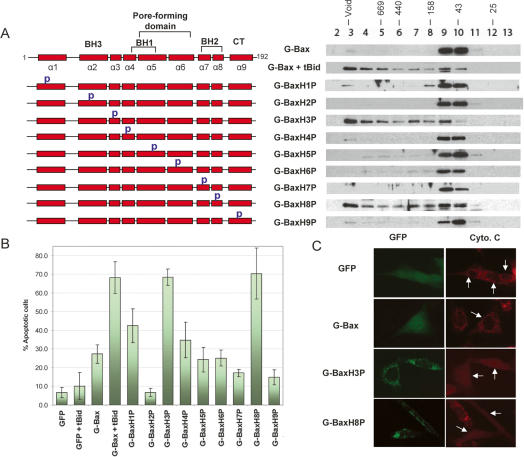
Figure 5.
Identification of constitutively active mutants of Bax. (A) Gel-filtration analysis of proline-scanning mutants of Bax in DKO cells. Schematic representation of the proline mutants of Bax are shown on the left. (p) The proline mutation. The amino acid residues for each proline mutant are specified in Materials and Methods. DKO cells were transfected with the expression plasmids of indicated wild type and proline mutants of Bax. All transfections in this figure were carried out in the absence of the tBid expression plasmid unless specified otherwise. (B) Apoptotic activities of the proline mutants of Bax in DKO cells. (C) Cytochrome c release from mitochondria induced by two proline mutants. Cells that were transfected with the indicated expression plasmids for GFP fusion proteins are shown on the left. Cells stained with cytochrome c antibody are shown on the right. Arrows point to the cells that express the indicated GFP fusion proteins.

