Fig. 1.
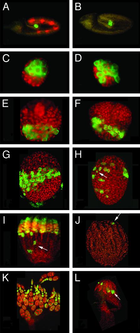
Germ cells degenerate starting at the late larva stage in γTub23CPI fs(2)TW11 double mutant females. Germ cells in control γTub23CPI fs(2)TW11/+ heterozygous animals (A, C, E, G, I, and K) and γTub23CPI fs(2)TW11 homozygous animals (B, D, F, H, J, and L) at different stages of development visualized with anti-Vasa antibody (green) and counterstained with anti-β-galactosidase antibodies (red) for a blue balancer (see Materials and Methods)(A and B) or the DNA stain TOTO-3 (red) (C-L). The stages shown are embryos (A and B) and ovaries from second-instar larva (C and D), third-instar larva (E and F), P1 pupa (G and H), P5/6 pupa (I and J), and 1-day-old adult (K and L). All gonads are oriented with their posterior end to the bottom. (A-D) We did not detect any difference between control and homozygous individuals from embryogenesis until second-instar larva. In mutant third-instar larva ovaries (F) the number of germ cells is slightly reduced in comparison to the WT (E; diameter of the gonad is 60 μm). Prepupa γTub23CPI fs(2)TW11 homozygous ovaries (H) contain fewer germ cells than control ovaries (G) and germ cells are dispersed throughout the gonad. In older pupae mutant ovaries contain only a few Vasa-positive cells (J), sometimes clustered (arrow). At this stage most WT germ cells are arranged within ovarioles (I) that already contain egg chambers and few clusters of germ cells lagging behind at the posterior half of the ovary (arrow). In WT adult ovaries (K) Vasa is present in the cysts in the germarium and is enriched in the nurse cells of later cysts. In γTub23CPI fs(2)TW11 double mutant ovaries (L) a few Vasa-positive cells and clusters are still present (arrow). (A-F, K, and L) Images were produced by conventional fluorescence microscopy. (G-J) Projections of confocal sections.

