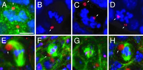
Fig. 2.
Mitotic divisions are affected in γTub23CPI fs(2)TW11 third-instar larva ovaries. Mitotic cells of the germ line in WT (A and E) and γTub23CPI fs(2)TW11 (B-D and F-H) third-instar larva ovaries. The germ cells were identified by immunostaining with an anti-Vasa antibody in A-D (green, shown only in A) or with anti-β-spectrin antibody in E-H (red). DNA is in blue in A-H, centrosomes in A-D were stained with an anti-centrosomin antibody (red), and microtubules in E-H were stained with anti-α-tubulin antibodies (green). (A-D) Centrosomes are positioned at opposite sides of the condensed chromosomes in a WT division (A, arrows). In mutant germ cells centrosomes are not correctly aligned (C and D, arrows). (B) A mutant mitotic cell with a single centrosomal signal (arrow). (E-H) Normal mitotic division in a WT germ cell (E). The spectrosome is associated with one spindle pole (arrow). In the mutant the spindle can be almost normal (H)to very poorly organized (F and G). The spectrosome is associated with the spindle, but not always with the spindle pole (F, arrow). All panels are projections of confocal sections. (Scale bar is 5 μm.)

