Fig. 3.
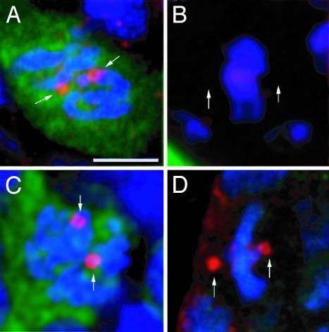
Cell type-dependent expression of γTub37C. Germ (A and C) and somatic (B and D) cells from WT third-instar larva ovaries stained (red) with antibodies that recognize only γTub37C (A and B) or both γ-tubulin isoforms (C and D). The germ cells are identified by immunostaining with an anti-VASA antibody (green), and DNA was stained with TOTO-3 (blue). γTub37C is detected at the centrosomes of dividing germ cells (A, arrows) but not in dividing somatic cells (B, arrows point to putative position of the centrososmes). All images are projections of confocal sections. The reddish appearance of DNA in B is caused by bleed-through of TOTO-3 into the red channel in this preparation and does not represent a specific signal. (Scale bar is 5 μm.)

