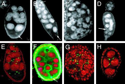
Fig. 5.
Egg chambers with incorrect number of germ cells in γTub23CPI fs(2)TW1RU34 mutant females. Immunofluorescence of egg chambers from WT (A and E) and γTub23CPI fs(2)TW1RU34 (B-D and F-H) females, stained with DAPI (gray in A-D and red in E-H). Ring canals are green in E, G, and H. Posterior is to the bottom. (B and F) Egg chambers produced by fusion of adjacent egg chambers. Two oocyte nuclei are located at opposite ends (arrows), and the total number of nurse cells corresponds to 30. (F) Tubulin staining (green) and nodkhc-LacZ localization (blue) reveal that an MTOC is associated with each of the two oocyte nuclei. (C and G) Two egg chambers containing >16 germ cells, likely produced by extra rounds of mitotic divisions judging by the nurse cell size, the shape of the egg chamber, and the mixed population of ring canals in G.(D and H) Shown are <16 egg chambers. There are follicle cells within the egg chamber in D (arrow). The egg chamber in H contains only four germ cells and three ring canals, suggesting an early arrest of the mitotic program. Images A-D were obtained by conventional fluorescence microscopy. Images E-H are projections of confocal sections. Egg chamber size is ≈150 μm.

