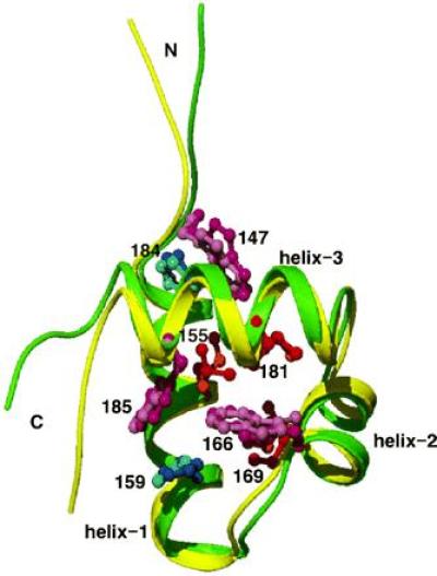
Figure 2.

The refined average structure of the 20 I155L mutant structures determined by NMR-distance geometry method. The backbone is shown in green. Side-chain atoms of Trp and His residues are shown in purple and dark blue, respectively. Side-chains of Leu-155, Ile-169, and Ile-181 are shown in red. The backbone of the superimposed wild-type structure (21) is shown in yellow, and side chains of Ile, Trp and His are shown in orange, pink, and light blue, respectively. The characters N and C indicate the N and C termini, respectively, and several residue numbers are indicated. The figure was drawn with the program raster3d (34). The coordinates for the average and the 20 I155L mutant structures were submitted to the Protein Data Bank.
