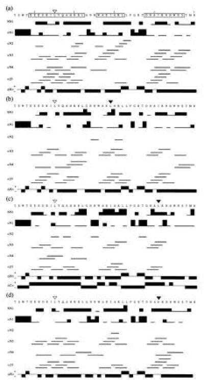
Figure 3.

Diagram of the sequential and short-range NOEs and the chemical shift indexes (36) observed for the mutant R3s from positions 145 to 190: I155L (a), I155L/I169L (b), I155L/I181L (c), and I155L/I181V (d). The abbreviations NN1, αN1, αN2, αN3, αN4, and αβ3 represent dNN(i, i+1), dαN(i, i+1), dαN(i, i+2), dαN(i, i+3), dαN(i, i+4), and dαβ(i, i+3), respectively. The thick, medium, and thin bars correspond to strong, medium, and weak NOEs, respectively. The downfield shifted 1Hα chemical shifts (negative ΔHα) and the upfield-shifted 13Cα chemical shifts (positive ΔCα) from the corresponding chemical shifts of a random coil are indicative of helical regions (35, 36). Helical regions in the amino acid sequence of I155L from the tertiary structure are boxed. Position 155 is indicated by an open triangle. Other substituted residues in the mutant R3s are indicated by filled triangles.
