Fig. 3.
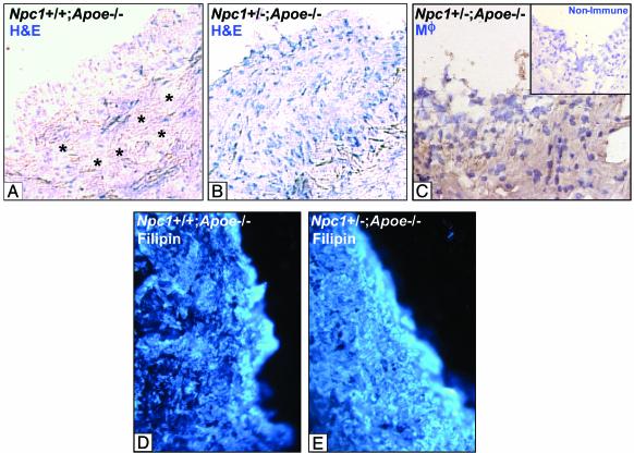
Hematoxylin, macrophage, and filipin staining of proximal aortic lesion sections from male Npc1+/+;Apoe-/- and Npc1+/-;Apoe-/- mice. (A and B) Sections of proximal aortic lesions from 25-week-old cholesterol-fed male Npc1+/+;Apoe-/- (A) and Npc1+/-;Apoe-/- mice (B). Sections were stained with hematoxylin and eosin (H&E). (×225.) Asterisks indicate acellular areas. The lesion from the Npc1+/-;Apoe-/- mouse (B) is markedly more cellular than the lesion from the Npc1+/+;Apoe-/- mouse (A). (C) These cells were identified as macrophages (Mϕ) by immunohistochemistry. Inset shows a nonimmune control. (D and E) Lesions from these mice were stained with filipin. Overall lesional accumulation of FC was not markedly different in the two lesions.

