Fig. 5.
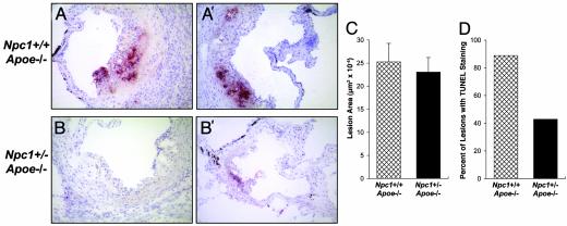
TUNEL staining of lesions in the proximal aortae of Npc1+/-;Apoe-/- and Npc1+/-;Apoe-/- mice. Atherosclerotic lesions in the proximal aortae of nine 18-week-old cholesterol-fed Npc1+/+;Apoe-/- mice and seven Npc1+/-;Apoe-/- mice were assayed for TUNEL positivity. Examples of representative sections from two Npc1+/+;Apoe-/- mice (A and A′) and two Npc1+/-;Apoe-/- mice (B and B′) are shown. (×20.) Quantification of total lesion area and the percentage of mice that had TUNEL-positive lesions are shown in C and D, respectively. For the TUNEL analysis, two aortic sections were examined per mouse. Using the Mann-Whitney U test, we found that the difference in lesion area between the two groups of mice was not statistically significant, whereas the difference in TUNEL positivity was found to be statistically significant by the χ2 test (P < 0.05).

