Figure 1.
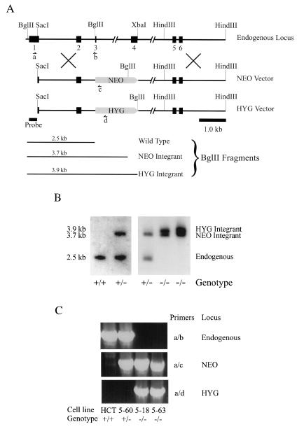
Targeted disruption of the human Smad4 gene. (A) Alignment of endogenous Smad4 locus with the two targeting vectors. Solid boxes represent exons 1–6. Drug markers are shown as shaded boxes with pointed ends indicating the simian virus 40 polyadenylation signals. Also shown are the locations of the PCR primers and the probe used for Southern blot analysis. The expected BglII restriction fragments hybridizing to the probe are diagrammed at the bottom. As a result of recombination between the vector and endogenous locus, exons 3 and 4 are entirely deleted. (B) Southern blots of BglII-digested genomic DNA. Genomic DNA was prepared from parental (+/+), heterozygote (+/−), and Smad4-null (−/−) HCT116 clones. After digestion with BglII and agarose gel electrophoresis, DNA was blotted and hybridized with the probe mapped in A, corresponding to sequences outside the 5′ homologous arm. (C) Genomic PCR of the clones used in this report. Genomic DNA was prepared from parental HCT116 cells (HCT) (+/+), and clones 5–60 (+/−), 5–18 (−/−), and 5–63 (−/−). Primer pairs (see A for positions) were specific for the deleted portion of endogenous Smad4 gene (Top), the NEO-targeted locus (Middle), or the HYG-targeted locus (Bottom).

