Figure 4.
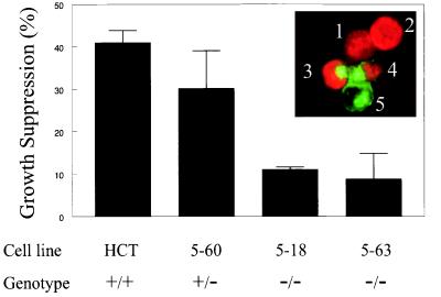
Effect of Smad4 on growth suppression by TGF-β. Cells were transfected with a vector encoding αGT plus TβRII (pαGT-RII) or an identical vector without TβRII (pαGT). The cells were then cultured in medium containing TGF-β1 (1 ng/ml), and after 36 hr, BrdUrd was added for an additional 3 hr. The cells were harvested, bound to fluorescein-labeled lectin, and stained with anti-BrdUrd antibodies. (Inset) Examples of Smad4−/− cells transfected with the TβRII vector pαGT-RII. The green fluorescence at the cell periphery and in the Golgi apparatus indicates expression of αGT. The red nuclear fluorescence indicates DNA synthesis. Cells 1 and 2 were not transfected but were synthesizing DNA. Cells 3 and 4 were transfected and were synthesizing DNA, whereas cell 5 was transfected but was not synthesizing DNA. In the graph, the fraction of BrdUrd-positive cells among pαGT-RII transfectants was normalized to the fraction of BrdUrd-positive cells in the control pαGT transfectants. Bars and brackets represent the means and standard deviations, respectively, determined from at least two independent assessments of 400 transfected (green) cells from a single experiment; similar results were obtained in two other independent transfections. All determinations were performed in a blinded manner. BrdUrd incorporation in pαGT-RII-transfected Smad4−/− cells was not statistically different from pαGT transfected cells, but the differences between the Smad4−/− homozygotes and parental cells was statistically significant (P < 0.05, Student’s t test).

