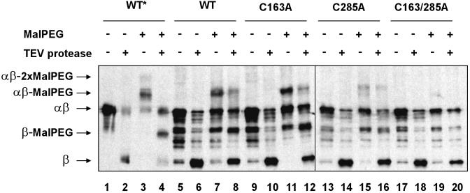
Fig. 5.
Alkylation of exposed cysteines on vesicles. Vesicles from FED126 cells expressing the designated constructs were prepared as indicated in Methods. Vesicles were incubated with TEV protease and malPEG as depicted by symbols + and -. Proteins were visualized by Western blotting using antibodies that react against the c-Myc epitope. The following plasmids were used: pFK269 (lanes 1-8), pFK270 (lanes 9-12), pFK271 (lanes 13-16), and pFK272 (lanes 17-20). The asterisk indicates that whole TCA-precipitated cells were used, instead of vesicles. Thus, bands on lanes 1-4 show the electrophoretic mobility for each of the possible species.

