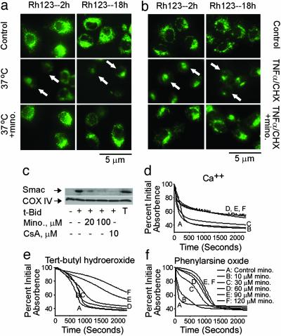
Fig. 4.
Minocycline inhibits dissipation of Δψm, release of Smac, and induction of PT. (a and b) Mutant huntingtin-expressing ST14 A cells were shifted at the nonpermissive temperature of 37°C in SDM (a), or ST14 A cells were treated with 10 ng/ml TNF-α/10 μM CHX (b) with or without 10 μM minocycline for 2 and 18 h. Cells were stained directly with 2 μM rhodamine 123. Arrows show dissipation of Δψm. (c) Purified mouse brain mitochondria were incubated with minocycline or cyclosporin A. Smac release was induced by tBid and inhibited by addition of minocycline or cyclosporin A. T, total mitochondrial Smac; CsA, cyclosporin A. With the mitochondrial pellets, cytochrome c oxidase subunit IV (COX IV) was used as a loading control. (d-f) Minocycline has distinct mechanisms of action against oxidative and nonoxidative PT inducers. Representative traces from a minimum of three experiments on the effects of minocycline on PT induction. Inducers: 50 μM Ca/Pi (d); 1 mM tert-butyl hydroperoxide 2 μMCa2+ (e); 50 μM phenylarsine oxide (f). PT induction was studied in liver mitochondria isolated from 4- to 6-month-old male Fischer 344 rats. Data for all traces in a single panel were collected at the same time from the same mitochondrial preparation.

