
Figure 4.
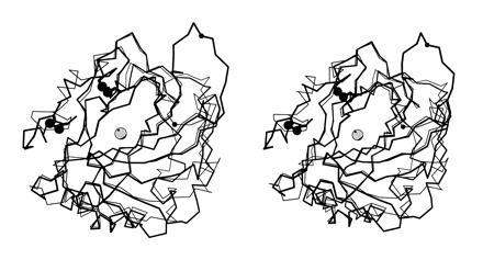
Least-squares Cα superposition of CA IV (thick bonds) and CA II (thin bonds). The active site zinc ion appears as a gray sphere, and disulfide linkages of CA IV are indicated by large black spheres. Note that although the overall folds of the two isozymes are generally similar, there are significant differences in the region of residue 131 (small black sphere). The extended conformation of the Arg-129–Asn-130 linkage in CA IV may enhance its susceptibility to proteolysis (17, 23).