
Figure 2.
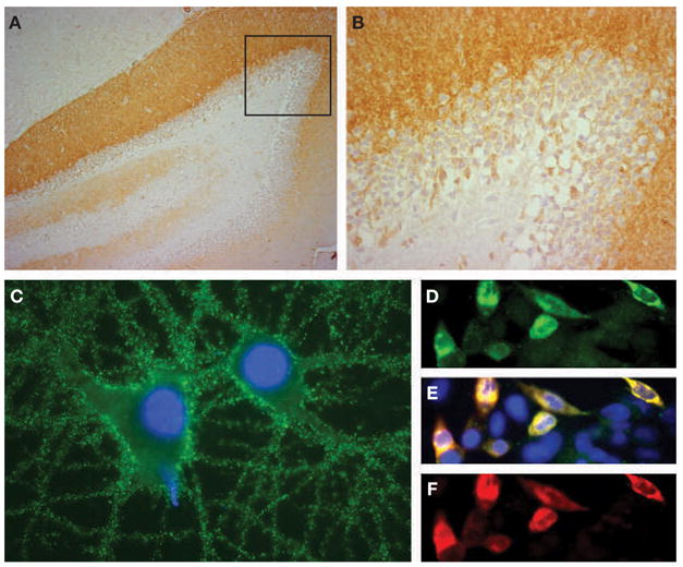
Demonstration of N-methyl-D-aspartate receptor antibodies in the patient’s cerebrospinal fluid. (A,B) Sagittal section of rat hippocampus incubated with the patient’s cerebrospinal fluid (CSF; diluted 1:20). The anti-N-methyl-D-aspartate receptor (NMDAR) antibodies produce intense immunolabeling of the molecular layer, adjacent to the granular cells of the dentate gyrus (faintly counterstained with hematoxylin). The area of the dentate gyrus included in the box is shown magnified in panel B. (C) Cultured rat hippocampal neurons incubated with the patient’s CSF demonstrate intense immunolabeling of NMDARs contained in the surface of neurons and neuronal processes (nuclei demonstrated by staining with DAPI). (D–F) Confirmation that the CSF antibodies selectively react with NMDARs is shown using human embryonic kidney (HEK293) cells expressing NR1/NR2B heteromers of the NMDAR. The reactivity of the patient’s antibodies (green, panel D) co-localize (yellow, panel E) with the reactivity of NR2B-specific antibodies (red, panel F). In addition, the patient’s antibodies reacted with NR1/NR2 heteromers containing NR2A, NR2C and NR2D subunits of the NMDAR, which have substantial homology with NR2B (not shown). All immunohistochemical techniques have been reported previously.2 Immunoperoxidase method was used in panels A (100×) and B (400×), and immunofluorescence in panels C (800×) and D–F (400×).