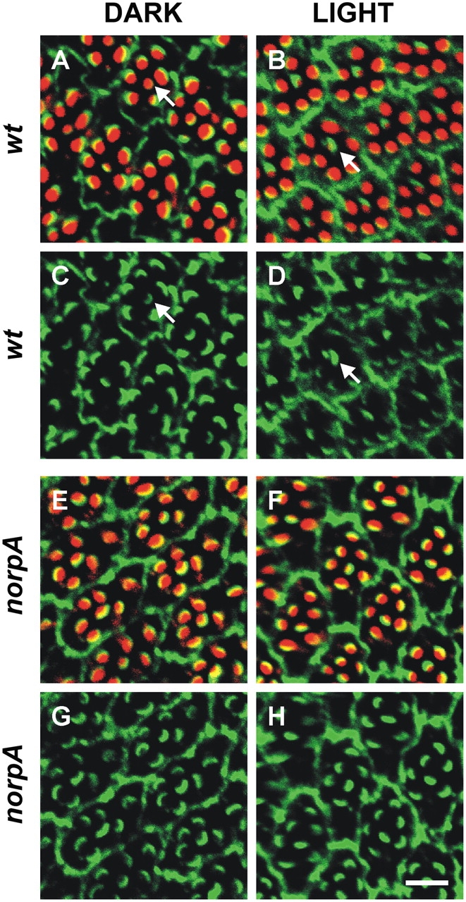
Figure 2.

Light-dependent movement of Dmoesin in the photoreceptor cell of WT D. melanogaster retina. (A–D) Cross sections through dark-raised and illuminated wt flies (Schott BG28 blue light for 1 h). Flies were double labeled with rhodamin-coupled phalloidin (red) and αDmoesin (1:2,000 dilution). Primary Dmoesin antibody was detected by a Cy5-coupled secondary antibody (green; C and D). The overlay of both markers appears yellow in some ommatidia (A and B). Arrows indicate R7 cells. (E and F) The same experiment as in A–D was performed with norpA P24 mutant. Bar, 8 μm.
