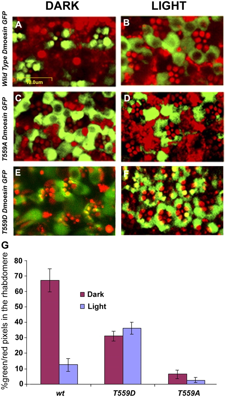
Figure 7.

Light- and phosphorylation-dependent movement of Dmoesin from the rhabdomere to the cell body. (A–F) Intracellular distribution of Dmoesin-GFP protein fusions, as observed in confocal micrograph cross sections of living D. melanogaster retinae of the transgenic lines: UAS Dmoesin-WT-GFP (A and B), UAS Dmoesin-T559A -GFP (C and D), and UAS Dmoesin-T559D-GFP (E and F). Green indicates GFP fluorescence. The strong autofluorescence of the rhabdomeres (red) allows localizing Dmoesin distribution with respect to photoreceptor compartments. Dark-raised flies were kept in obscurity (A, C, and E) or submitted to blue light illumination (B, D, and F). In flies expressing Dmoesin-WT-GFP (A and B), the fluorescent protein moves from the rhabdomere and cortical actin regions to the cell body of photoreceptors in response to light. Almost all Dmoesin-T559A-GFP (C and D) proteins accumulate outside of the rhabdomeres, independently of the illumination regime, and a significant fraction of Dmoesin-T559D-GFP (E and F) was observed in the rhabdomeres and cortical actin regions regardless of illumination regime. (G) The histogram plots the ratio of the number of green (GFP) to red (autofluorescence) pixels in the rhabdomere and cortical actin regions, as defined by the area that displays autofluorescence. P < 0.01; n = 20 for each fly strain. The error bars are SEM.
