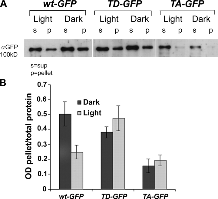
Figure 8.
The light-dependent movement of Dmoesin from the membrane to the cytosol is blocked in the Dmoesin-T559A and Dmoesin-T559D mutants. (A) Western blot analysis of Dmoesin distribution in membrane-bound or -soluble fractions of D. melanogaster head protein extracts. Membrane and soluble proteins extracted from dark-raised and illuminated Dmoesin-GFP transgenic lines were Western blotted using αGFP. Extracts were prepared from the same fly strains as Fig. 7, as indicated. (B) The histogram plots the ratio of membrane-bound to total Dmoesin signals from replicate experiments similar to that shown in A. Although illumination halves levels of WT Dmoesin in association with membranes (P < 0.01; n = 3), no significant modification of Dmoesin distribution is provoked by illumination of Dmoesin-T559A-GFP and Dmoesin-T559D-GFP. The error bars are SEM.

