Figure 3.
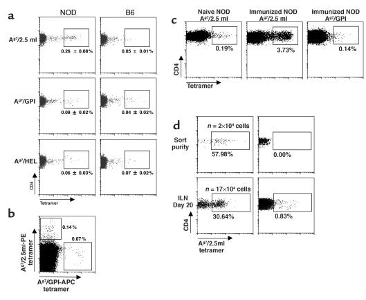
Presence of 2.5mi+ T cells in NOD mice. (a) Specific 2.5mi+CD4+ T cells were identified in subcutaneous LNs from young adult NOD but not from B6 mice. Control stains for either Ag7/GPI or I-Ag7/HEL tetramers are shown in the lower panels. (b) Lack of cross-reactivity of Ag7/2.5mi tetramers with nonspecific T cells. Splenocytes from a NOD mouse were stained with PE-labeled Ag7/2.5mi tetramers, followed by incubation with APC-labeled Ag7/GPI tetramers. T cells were only stained by either of the tetramers, not by both simultaneously, indicating specific recognition. (c) 2.5mi+CD4+ T cells expansion after immunization with the 2.5 mi+ peptide. Popliteal LN cells from nonimmunized and immunized NOD mice were stained with either Ag7/2.5mi or Ag7/GPI tetramers. (d) Expansion of 2.5mi+CD4+ cells in a lymphopenic mouse reconstituted with 2.5mi+CD4+ cells (or 2.5mi–CD4+ cells as control), sorted from splenocytes of 5-week-old NOD mice and immediately analyzed for sort purity. Twenty thousand sorted cells were injected into RAGo/o/NOD mice. Inguinal LN cells were stained 20 days after transfer for the presence of 2.5mi+CD4+ cells. The number of injected cells was not corrected for the presence of nonviable cells and is therefore likely to be overestimated. The number of 2.5mi+CD4+ cells present in the whole mouse (n) was extrapolated to a full lymphoid compartment from the number of 2.5mi+CD4+ cells observed in the inguinal LN by flow cytometry (taking into account multiple losses during the staining procedures and the flow cytometry acquisition itself).

