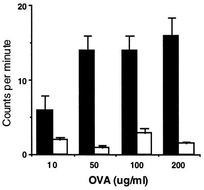
Figure 4.
Proliferation of spleen cells from mice treated with 1 mg OVA at birth and immunized with OVA (50 μg)/alhydrogel at 4 weeks of age then subsequently aerosol challenged 12 days later. Spleen cells were stimulated in vitro with OVA for 72 h and then 3H incorporation determined. OVA-specific T cell proliferation was determined as incorporation levels (cpm) above background and normalized to incorporation levels from anti-CD3 stimulated cells. Splenocytes from mice treated neonatally with OVA (open bars) show significantly reduced proliferative ability at all concentrations of OVA compared with splenocytes from PBS-treated control mice (P < 0.05, n = 3).

