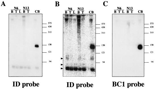
FIG. 3.
Northern blot analysis of RNAs extracted from different tissues of BC1-deficient mice. (A) Hybridization with a probe complementary to the 5′ ID domain of BC1 RNA (SB358). Lanes B, T, and L, RNA samples extracted from brain, testes, and liver, respectively, of homozygous BC1-deficient mouse lines 6 and 13. Lane CB, RNA extracted from brains of control mice which exhibits BC1 RNA of ∼150 nt. The size marker positions are shown in nucleotides on the right. (B) After exposure on a phosphorimager, additional hybridization signals corresponding to RNAs migrating faster than BC1 RNA are observed. Arrowheads identify signals at ∼110, ∼90, and ∼75 nt. (C) Hybridization with a probe complementary to the 3′ unique domain of BC1 RNA (probe 90).

