
Figure 2.
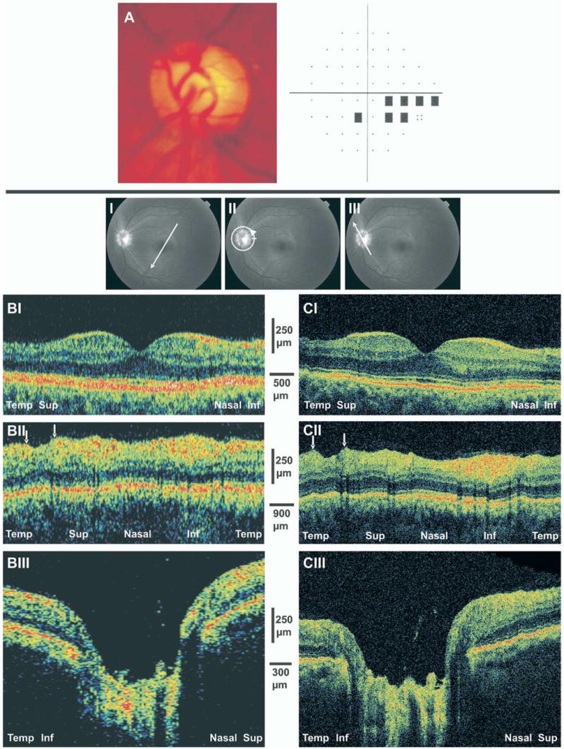
A, Optic disc photograph of early glaucomatous damage demonstrating moderate cupping and superior temporal neuroretinal rim thinning. A visual field defect is presented in the inferior nasal region. B, StratusOCT scans. C, Ultrahigh-resolution optical coherence tomography images. I, Thinning of the nerve fiber layer (NFL) and ganglion cell layer is evident in the temporal superior macular scans. II, Arrows highlight focal NFL defects in the peripapillary scans. III, Shallow cupping is evident in the optic nerve head scans. Inf = inferior; Sup = superior; Temp = temporal.