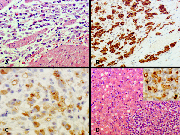
Figure 3.
An invasive adenocarcinoma is present in the gastrectomy specimen from patient 1. Numerous signet ring cells are seen in the gastric wall (panel A). Carcinoma cells are immunohistochemically positive for CK7 (panel B) and gross cystic disease fluid protein (GCDFP) (panel C). Metastatic carcinoma cells in the lymph node (panel D) are also positive for GCDFP (insert).

