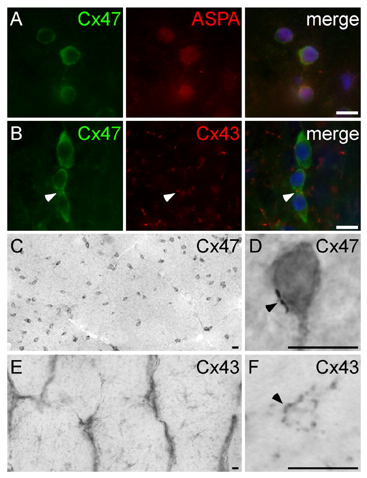
Figure 7. Primate oligodendrocytes express Cx47.

These are images of sections of rhesus monkey optic nerve, immunostained with mouse monoclonal antibodies against Cx47 (A–D) or Cx43 (E–F), a rabbit antisera against aspartoacylase (ASPA; A) or Cx43 (B), visualized with FITC- or TRITC- (A&B) or peroxidase-conjugated secondary antiserum (C–F), and counterstained with DAPI (A&B, merged panels). Cx47-immunoreactivity is diffusely dispersed within small cell bodies (A–B, green; C–D), with occasional gap junction plaques (arrowhead; D), and colocalizes with ASPA (A), a marker of oligodendrocytes. Cx43-immunoreactivity is found on astrocyte cell bodies and their proximal processes (E) and in gap junction plaques that are distributed throughout the optic nerve, including on small cell bodies that are likely to be oligodendrocytes (arrowheads, B&F). Scale bars: 10μm.
