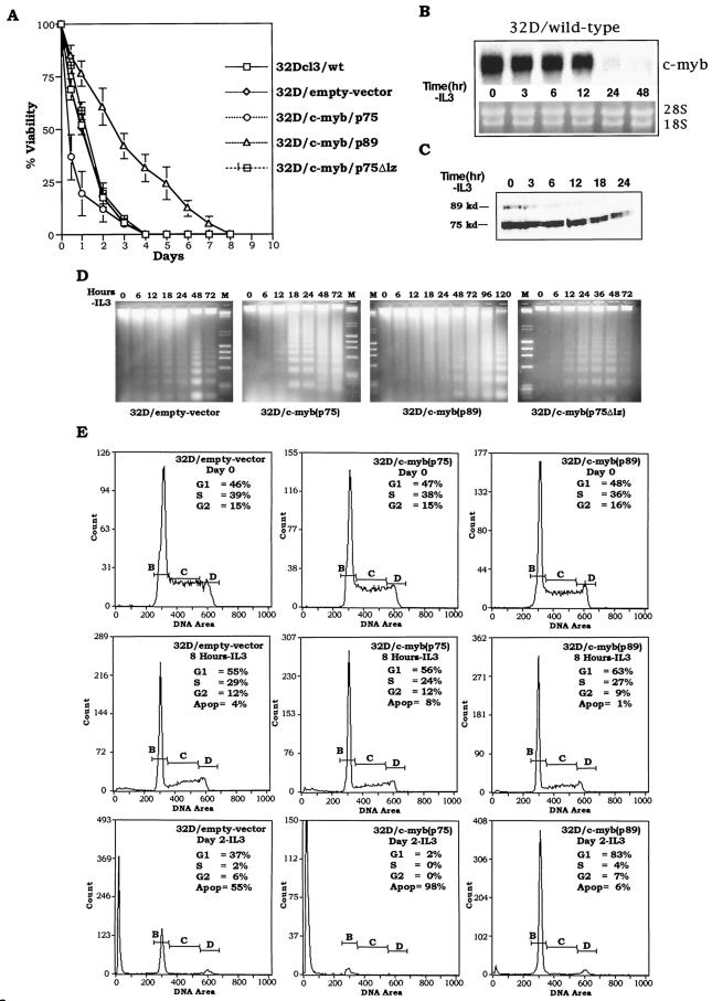
FIG.3.
Effect of p75, p75Δlz, and p89 c-Myb proteins on IL-3 withdrawal-induced apoptosis of 32Dcl3 cells. (A) Wild-type, mock-transfected (empty-vector pMTneo) and 32Dcl3 cells expressing exogenous p75, p75Δlz, and p89 c-Myb proteins were washed in IL-3-free medium and incubated up to 8 days. At each indicated time point, the cells were analyzed for viability by trypan blue exclusion. The curves represent a mean of three experiments. (B and C) Wild-type 32D cells were subjected to IL-3 withdrawal, and the quantity of c-myb mRNA and protein was measured at the indicated time points by Northern blot and Western blot analyses, respectively. As a control for RNA loading, filters were stained with ethidium bromide to compare the levels of 28S and 18S RNAs. (D) Analysis of DNA fragmentation. At the indicated times following IL-3 withdrawal, DNA fragments released from 10 × 106 cells from different 32D cell lines were extracted, separated by electrophoresis, and stained with ethidium bromide. (E) Cell cycle analysis of empty-vector, p75c-myb and p89c-myb cells at day 0, 8 h, and day 2 following IL-3 withdrawal. The cells were fixed and stained with propidium iodide, and DNA content was measured with a flow cytometer. The horizontal lines designated B, C, and D in the graphs represent the amount of DNA in the cells (1N, intermediate, and 2N, respectively) and therefore correspond to cells in G1, S, and G2 phases, respectively.

