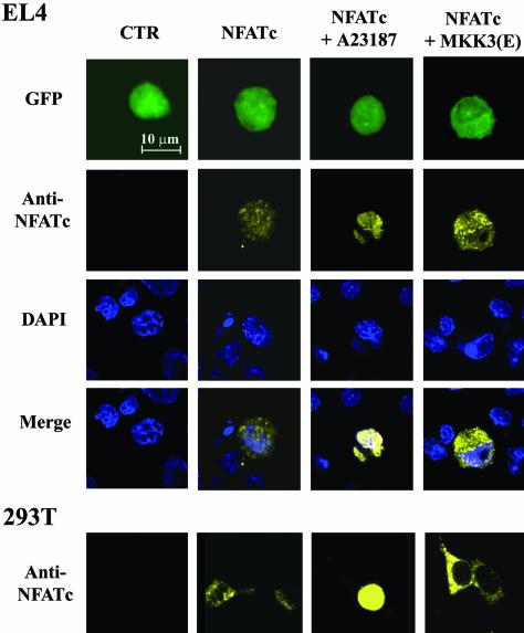
FIG. 4.
Confocal image of intracellular distribution of NFATc stimulated with MKK3(E) or A23187. EL4 T cells and 293T cells were transfected with EGFP only (CTR [control]); EGFP and CMV-NFATc (NFATc); EGFP, CMV-NFATc, and A23187 (NFATc + A23187); and EGFP, CMV-NFATc, and MKK3b(E) [NFATc + MKK3(E)]. At 24 h after transfection, cells were fixed with 3.7% paraformaldehyde, followed by methanol permeabilization. The cells were stained with DAPI (DAPI) and with anti-NFATc followed by phycoerythrin-conjugated anti-mouse immunoglobulin (anti-NFATc). The NFATc expression of cells was analyzed by use of Zeiss confocal laser scanning microscope LSM 510 with a ×63 objective lens. Green cells (GFP) indicate transfected cells, while DAPI indicates the nucleus. The nuclear localization of NFATc was examined by overlapping the anti-NFATc-stained image with the DAPI-stained image (merge).

