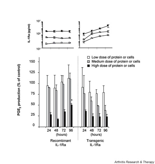
Figure 3.

Comparison of the relative inhibitory activity of HIG-82-IL-1Ra+ cells to recombinant IL-1Ra following extended incubation. HSF cultures were incubated with one of three doses of rIL-1Ra (3, 30, or 300 ng/ml) or IL-1Ra producing cells (3 × 10³, 2 × 10⁴, or 1 × 10⁵) that secreted corresponding levels of transgenic IL-1Ra protein within 24 hours. From the results of Fig. 2 these doses provided either low (approximately 10–15%), medium (approximately 25–50%), or high level (approximately 70–80%) inhibition of IL-1β. Twenty-four hours following the addition of the source of IL-1Ra, 5 ng recombinant IL-1β was added to each culture well. At 24 hour intervals after IL-1 stimulation, PGE2 and IL-1Ra levels in the conditioned media were measured. PGE2 levels were normalized to IL-1 stimulated HSF that were not treated with IL-1Ra, which were assigned the value of 100%. The bottom graph represents the change in PGE2 levels in the media over time from cells receiving either rIL-1Ra or the tIL-1Ra producing cells. The white bars represent cultures receiving the low dose of protein or cells, the grey bars the medium dose, and the black bars the high dose. The inset above reflects the corresponding IL-1Ra concentrations in the conditioned media for each time point and dose. Experiments were performed in triplicate, and each data point repesents the mean value ± SD. *P < 0.05 versus corresponding IL-1Ra source at 24 hours. HSF, human synovial fibroblast; IL-1Ra, IL-1 receptor antagonist; PGE2, prostaglandin E2.
