
FIG. 5.
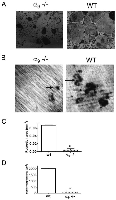
OCL formation and bone resorption in bone marrow cultures from α9−/− mice. Bone marrow cells from 7-day-old α 9−/− or WT mice were cultured with RANKL (50 ng/ml) and M-CSF (10 ng/ml) for 7 days. The cultures were fixed and stained for TRACP. (A) OCLs formed from α9−/− were smaller and more contracted than those in WT cultures. After 7 days, the cells were removed from dentin. The resorption pits formed on dentin were visualized by light microscopy after staining with hematoxylin. (B) α9−/− OCLs formed dramatically fewer resorption pits. In contrast, WT OCLs formed numerous and serpentine-like resorption pits. In addition, OCL resorption capacity was determined by measuring (C) total bone resorption areas and (D) the average resorption area per OCL. Magnification: ×10. Similar results were found in three independent experiments. Results from one typical experiment are shown as mean ± SE (*p < 0.05).