
FIG. 6.
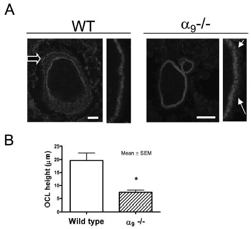
Cytoskeleton dysfunction and actin-ring deformation in α9−/− OCLs. (A) OCLs formed from α9−/− and WT marrow cultures were stained with Texas-red-phalloidin (magnification, ×10; scale: 25 μm). (A) Left: actin-ring formation in WT and α 9−/− OCL; fine structure of the podosome belt is shown at higher power. Open arrow shows the lamellipodium. Solid arrows show the loosely distributed podosomes at the edge of actin-ring. (B) Osteoclast height in WT and α9−/− OCLs. Similar results were found in three independent experiments. Results from one typical experiment are shown.