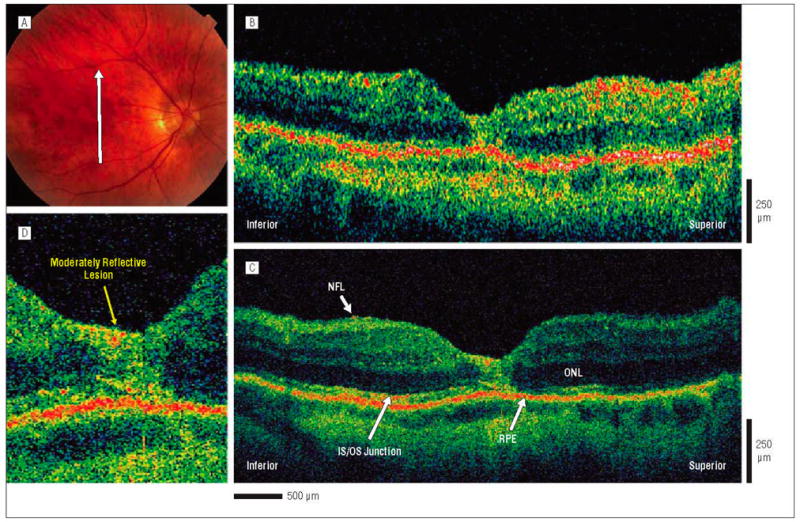
Figure 5.

Postoperative macular hole, 1 month after surgical repair. The patient is a 66-year-old man who underwent initial evaluation for a stage 2 macular hole in the right eye and had a preoperative visual acuity of 20/80. He underwent pars plana vitrectomy with internal limiting membrane peel and injection of 25% sulfur hexafluoride gas. One month later, the hole was flat and closed on results of examination, and visual acuity was 20/70. A, Fundus photograph shows the direction of optical coherence tomography (OCT) images. B, Standard-resolution OCT image acquired in the scan direction. The photoreceptor IS/OS junction appears disrupted, although it is difficult to distinguish this layer. C, Corresponding UHR-OCT image acquired in the scan direction. The intraretinal layers are better visualized in the UHR-OCT image, and it shows the large, moderately reflective foveal lesion replacing the normal foveal anatomy. D, Magnification (×2) of the UHR-OCT image. A large, moderately reflective lesion is replacing the normal foveal anatomy. All of the intraretinal layers of the fovea, including the photoreceptor inner and outer segments of the outer retina, appear to be involved. Abbreviations are described in the legend to Figure 1.
