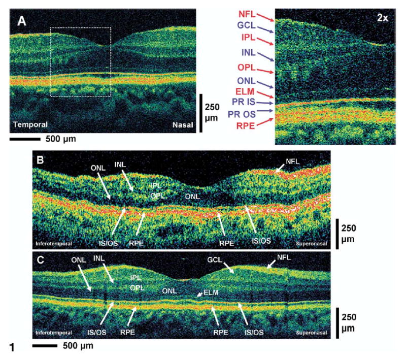
Figure 1.

A, High-magnification ultrahigh-resolution optical coherence tomography (UHR, OCT) image of the normal human macula. An enlargement of the image demonstrates the ability to visualize intraretinal layers that can be correlated with intraretinal anatomy: nerve fiber layer (NFL), ganglion cell layer (GCL), inner plexiform layer (IPL), inner nuclear layer (INL), outer plexiform layer (OPL), outer nuclear layer (ONL), external limiting membrane (ELM), photoreceptor inner and outer segments (PR IS, PR OS), retinal pigment epithelium (RPE). Red labels indicate high-backscattering layers, blue labels, low-backscattering layers. B, C, Standard StratusOCT and UHR OCT images, respectively, of the normal human macula. Most of the major intraretinal layers can be visualized in the StratusOCT image, but the GCL and ELM are much better visualized in the UHR OCT image.
